18:22
Oral lesion detection in seconds
AI-assisted screening for oral mucosal lesions. Take a photo, get a personalised clinical recommendation.
510(k)
Cleared
NHS
Validated
For screening purposes only · Not a medical diagnosis
‹ Back
Step 1 of 9
Before we begin
Please read the following information about how DentClair works and its limitations.
Your data & privacy
Photos are processed on-device and deleted immediately after analysis. No images are stored or transmitted to external servers.
Peer-reviewed clinical validation
Published in npj Digital Medicine (2024). Validated on 1,240 clinical cases. Sensitivity 89%, specificity 84%. Endorsed by NHS consultant oral medicine specialists.
Urgent symptoms
If you are experiencing severe pain, rapid swelling, or difficulty swallowing or breathing, please seek emergency care immediately.
‹ Back
Step 2 of 9
How to take a good photo
Image quality directly affects analysis accuracy. Follow these steps for the best result.
1
Good lighting is essential
Use natural light or a bright lamp. Avoid dark rooms. The oral cavity must be clearly visible.
2
Open wide and hold still
Use a mirror to position yourself. Keep the camera 15–20 cm from the lesion. Avoid blurry shots.
3
Focus on the lesion
Tap the lesion on screen to focus. The area of concern should fill at least 40% of the frame.
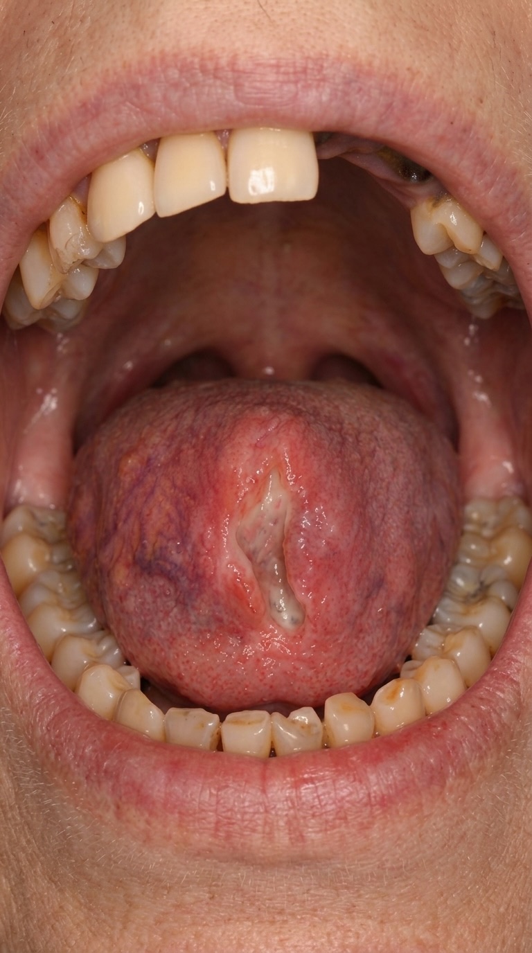
4
Take 2–3 photos
The app will automatically select the sharpest image for analysis.
DentClair
⚡ Auto
Position lesion inside the frame
‹ Back
Step 3 of 9
Image quality check
Your photo has been assessed for analysis suitability.
Sharpness
82%
Brightness
74%
Coverage
91%
Contrast
68%
Image is suitable for analysis. Proceeding with AI evaluation.
‹ Back
Step 5 of 9
Region detected
The AI has identified a region of interest. Please confirm this is the lesion you are concerned about.
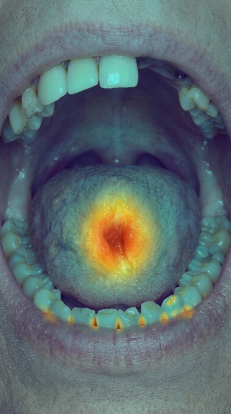
Detected characteristics
Irregular border
Mixed colouration
~8 mm diameter
Lateral tongue
Is this the lesion you are concerned about?
✓ Yes, that's it
No, retake photo
‹ Back
Step 6 of 9
Tobacco & alcohol exposure
These are the primary modifiable risk factors for oral mucosal lesions and oral cancer.
Tobacco use
Current smoker
Cigarettes, cigars, pipe — regular use
Smokeless tobacco
Chewing tobacco, snuff, betel quid, paan
Former smoker
Stopped more than 12 months ago
Never used tobacco
Duration of tobacco use
✓Less than 5 years
✓5–15 years
✓More than 15 years
✓N/A
Alcohol consumption
Daily or near-daily use
More than 14 units per week
Moderate use
1–14 units per week
Occasional or none
Less than weekly
Other relevant factors
✓Poor oral hygiene
✓Ill-fitting dentures
✓HPV history
✓Sun exposure (lip)
✓Immunosuppression
✓Family history of oral cancer
✓None of the above
Analysing your data
Please wait while the AI processes your information
15
seconds
‹ Back
Step 7 of 9
Assessment complete
The AI has finished analysing your data. Your clinical recommendation is ready.
Triage result
Urgent referral recommended
A potentially significant finding was identified. Please seek specialist review within 2 weeks.
Clinical recommendation
AI-assisted triage assessment · For discussion with your clinician
Triage classification
Urgent referral recommended
Seek specialist review within 2 weeks
Assessment summary
The identified lesion displays features associated with a potentially malignant disorder of the oral mucosa. Findings are consistent with erythroleukoplakia in the context of significant tobacco exposure. A definitive diagnosis requires clinical examination and histopathological analysis.
Findings → Probable diagnosis
Top diagnosis
68%
High risk
Diagnosis
Erythroleukoplakia
Potentially malignant disorder
Key findings
Irregular border with red-white surface variation
68%
Lateral tongue — high-risk anatomical site
22%
Tobacco exposure >15 years (primary risk factor)
10%
DentClair